Brain anatomy
This page is a dumping ground for anatomical terms that come up in conversation with neuroscientists, phrased (hopefully!) in a way that CS people can understand them and locate them in the brain. Click a tract name for more detailed information, including instructions for segmenting the tract out of a tractogram in Brainapp.
If terms like "posterior", "inferior", and "sagittal" are confusing to you, check out the Wikipedia articles on anatomical directions and anatomical planes.
Types of Tissue
The tissue of the nervous system may be grossly divided into two types: grey matter and white matter. In the grey matter, neuron bodies are packed relatively closely together, while the white matter is made up mostly of neuronal projections, called axons. The outer surfaces of the brain as well as a few internal structures are composed of grey matter, while much of the interior of the brain is composed of white matter. Bundles of axons in the white matter are called tracts and connect different grey matter regions together.
Parts of the Brain
The following diagram shows the lobes of the cerebrum, the outer structures of the forebrain.

The structure inferior to the temporal lobe and posterior to the spinal cord is the cerebellum.
The outer grey matter layer of the brain is called the cortex.
White Matter Tracts
-
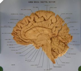 A sagittal section of a human brain. Click this image for a high-res version in which you can read the annotations.
 -
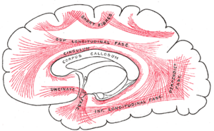 Major association fibers in the cerebrum, in a mid-sagittal lateral view.


There are three major types of tracts in the brain:
- Commissural fibers or transverse fibers connect equivalent grey matter sites in the two hemispheres of the brain.
- Association fibers connect different cortical grey matter sites to each other.
- Projection fibers connect cortical sites to subcortical grey matter or to the spinal cord.
Commissural Tracts
See [1] for an illustration of the comissural tracts of the brain.
![[1]](http://www.debratylermedart.com/medillus_webimages/commisuralfiberswebcopy.jpg)
- Corpus callosum (CC)
- The large tract of left-right-running fibers connecting the hemispheres. In a mid-sagittal cutting plane, it looks like a letter C turned on its side, with the tips pointing inferior; see [2]. Though the distal portions of the fibers that make up the CC are grouped into separate tracts in the interior of each hemisphere, they are collectively called the CC where they cross the mid-sagittal plane. In this sense the CC is more of a place name than a tract name; it indicates only the portions of those fibers that are near the mid-sagittal plane. Moving around the CC from the back (posterior), it is subdivided into parts named:
- Splenium
- The posterior, slightly bulbous part of the CC.
- Body
- The more-or-less flat part of the CC between the splenium and the genu.
- Genu
- The anterior part of the CC, where it bends back on itself. "Genu" means "knee".
- Rostrum
- The part of the CC inferior and posterior to the genu.
- Forceps major / forceps posterior
- The posterior projection of the CC (from the splenium) into the occipital lobe (the back of the brain). In an axial section, it looks like a letter C at the posterior portion of the brain with the tips pointing posterior. See [3].
- Forceps minor / forceps anterior
- The anterior projection of the CC (from the genu) into the cerebrum (the front of the brain). In an axial section, it looks like a letter C at the anterior portion of the brain with the tips pointing anterior. In other words, it looks sort of like a pair of mandibles over the eyes. See [4].
- Tapetum
- A medial projection of the CC (from the body) into the temporal lobe. In a well-reconstructed 3D tractogram, it forms a small horizontal fanning sheet of fibers emanating from the CC.
Association Tracts
- Cingulum / cingulum bundle
- White matter fibers located just superior to the corpus callosum, but running anterior-posterior. In a para-sagittal section, they look like a letter C turned on its side, with the tips pointing inferior, stacked right on top of the CC. In a coronal section, they look like two small circles just superior to the CC on either side of the interhemispheric fissure.
- Fornix
- C-shaped pathways that surrounds the subcortical gray matter in each hemisphere. It connects the hippocampus to the hypothalamus. Difficult to track due its curvature and partial voluming with gray matter.
- Inferior longitudinal fasciculus (ILF)
- A pathway that runs from the occipital to temporal cortices in each hemisphere. The majority of the fibers are in the temporal lobe.
- Superior longitudinal fasciculus (SLF)
- A collection of pathways that run between the frontal lobe and the temporal, parietal, and occipital lobes in each hemisphere. There are several subdivisions typically made based on their posterior connections.
- SLF I
- The portion of the SLF that intersects the corpus callosum and connects superior frontal cortex to parietal and occipital cortex. Typically not reconstructed with the single tensor model.
- SLF II
- The portion of the SLF that connects lateral frontal white matter (such as DLPFC) to the parietal and occipital cortices. Sometimes reconstructed with single tensors.
- SLF III
- The portion superior to the arcuate that connects lateral-inferior frontal cortex to parietal cortex. These fibers are very similar to the arcuate but do not curve.
- Arcuate fasciculus (AF)
- The portion of the SLF that curves to connect frontal cortex to the temporal lobe.
- Uncinate fasciculus
- A pathway that connects inferior frontal cortex to the temporal pole in each hemisphere. This curves around the insula cortex. Sometimes difficult to track due to high curvature.
Projection Tracts
- Corona radiata
- A fan-shaped sheet of WM that radiates superior (upward) from the brain stem to the cortex in each hemisphere. The fibers of the corona radiata run superior-inferior, and the fanning is anterior-posterior. The sheet is flat and thin in the left-right dimension; its orientation is approximated by a sagittal cutting plane.
- Corticospinal tract (CST)
- A major descending pathway that projects from the cerebral cortex to the spinal cord. It is the key pathway for voluntary movement (contrast it with the other major longitudinal but ascending pathway: the dorsal-column medial lemniscal system). Also, this is a good point to remember that tracts are generally named in source-to-target fashion. For example, corticospinal tract originates from the cortex and projects to the spinal cord, similarly corticobulbar tract goes from the cortex to the bulbar (well, brain stem), etc.
- Internal capsule
- White matter structure that runs between the cortex and medulla, containing major longitudinal (both ascending and descending) pathway systems, including the corticospinal tract, dorsal column medial lemniscus (DCML) system (only medial lemniscus part), corticobulbar tract, etc.
Grey Matter Structures
- Thalamus
- Major relay nuclei (grey matter structures) in the diencephalon (division of the brain primarily containing thalamus and hypothalamus in mature brain). Most sensory information is carried to the thalamus first (i.e., via ascending tracts) and then relayed to the cerebral cortex. Similarly, neural signals controlling movements, learning, memory and emotions bound to sub-cortical structures are relayed by the thalamus to their final destinations.
- Hippocampus
- Paired horn-shaped structures beneath the medial surface of the temporal lobes. Each half of the hippocampus is an elaboration of the edge of the cerebral cortex, where it folds up and inward from the inferior surface of the temporal lobe and curls around on itself. The hippocampus is associated with spatial memory.
External Links
- BrainNavigator --- an interactive 3D atlas application with labeled brain structures and histology thumbnails for rat and mouse brains. Does not include WM structures.
- BrainInfo --- an interactive visual and textual guide to thousands of named anatomical structures of the macaque brain. Includes WM regions (e.g. "inferior temporal white matter") but not fascicles ("superior longitudinal fasciculus").
- BrainMaps.org --- a database of histology of brains from various species, with an interactive online viewer
- WM Anatomy Tutorial with DTI pictures
- Biology-Online dictionary --- search for text descriptions of terms
- U. Arkansas neuroanatomy atlas images
- Radiopaedia --- a wiki encyclopedia for radiology; search for articles
- Gray's Anatomy --- an illustrated anatomy textbook from 1918
- MRI Studio is a program that supposedly includes Mori, et al.'s 2008 multi-subject DTI-based WM atlas