CS295J/Project Schedule:Steve: Difference between revisions
Steven Gomez (talk | contribs) |
Steven Gomez (talk | contribs) |
||
| Line 25: | Line 25: | ||
* A copy of all imaging data (from 40 subjects) used to build the [http://www.loni.ucla.edu/Atlases/Atlas_Detail.jsp?atlas_id=12 LONI Probabilistic Brain Atlas] is downloaded and available at: | * A copy of all imaging data (from 40 subjects) used to build the [http://www.loni.ucla.edu/Atlases/Atlas_Detail.jsp?atlas_id=12 LONI Probabilistic Brain Atlas] is downloaded and available at: | ||
/research/graphics/users/steveg/data/brain_atlases | /research/graphics/users/steveg/data/brain_atlases | ||
* '''STILL NEED''' -- MRIs with artifacts that make them "poor quality" | |||
== Evaluation Protocol == | == Evaluation Protocol == | ||
Revision as of 16:05, 8 November 2011
How do analysts assess scan quality in MRI imaging? How does that inform tool design?
Project Schedule
Nov 1 -- Filling out the paper: Abstract, related work and intro drafted; outlines for results and methods. Update on data and contact with Win.
Nov 8 -- Test MRIs (hopefully 10) collected and organized; evaluation protocol completed
Nov 15 -- Video captures of research assistants analyzing MRIs for QA (first session)
Nov 22 -- continued: Video captures of research assistants analyzing MRIs for QA (second session)
Nov 29 -- Coding of video captures completed; will share findings
Dec 6 -- Analysis of codings; figures for final paper created
Dec 13 -- Final draft, two-page extended abstract of research project
Report Drafts
- media:6nov2011-cs295j-report-steveg.pdf, 19:13, 6 November 2011 (EST)
Data and Resource Links
- A copy of all imaging data (from 40 subjects) used to build the LONI Probabilistic Brain Atlas is downloaded and available at:
/research/graphics/users/steveg/data/brain_atlases
- STILL NEED -- MRIs with artifacts that make them "poor quality"
Evaluation Protocol
Subjects: 4 research assistants in Win Gongvatana's group who have been previously trained in assessing scan quality.
Data: Each subject will receive a set of 10 T1-weighted MRI scans that have been shuffled in random order. 5 scans will be "normal quality" and taken from the component scans used to build UCLA's LONI Probabilistic Brain Atlas. The other 5 scans will contain artifacts that make them "poor quality". All will be of normal, human brains.
Method: We will use a think-aloud protocol and ask subjects to examine and score all scans in one session. Subjects will use whichever viewing tools they regularly use to assess scan quality. The observer will prompt each subject to explain what s/he is looking for in each image. Video of the subject will be recorded during this process, and the screen will be captured in order to do a post-hoc analysis of time spent per frame.
Questionnaire: After completing the tasks, subjects will be given a questionnaire with the following questions:
- High-quality images
- Please describe the properties or features of a scan that you would typically consider "high quality".
- Are these features obvious or subtle?
- How do you go about visually finding them?
- Low-quality images
- Please describe the properties or features of a scan that you would typically consider "poor quality".
- Are these features obvious or subtle?
- How do you go about visually finding them?
- Current QA process
- How much time do you typically spend doing quality checks on an MRI scan?
- How many slices do you typically look at during this process? (Please give both the number and the percentage of total, if not all are examined.)
- Please estimate the ratio of "poor quality" scans to "high quality" scans you come across in your research.
- Improving the process
- Do you wish the QA process were faster to complete?
- Do you wish the QA process were easier to complete?
- What tools, real or imaginary, would you use to help your QA process if they were available?
Boards
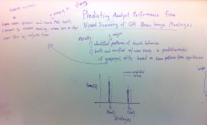
- Poster sketches and in-class critiques
-
 Oct 20, 2011
 -
 Oct 18, 2011
 -
 Oct 13, 2011


