New page: Here are some loose guidelines for placing the selection boxes to capture the corticospinal tract (CST). # Turn on the b=0 image in mid-sagittal view. # In one hemisphere, place a rectan...
(One intermediate revision by the same user not shown)
Line 1:
Line 1:
Here are some loose guidelines for placing the selection boxes to capture the corticospinal tract (CST).
Here are some loose guidelines for placing the selection boxes to capture the corticospinal tract (CST).
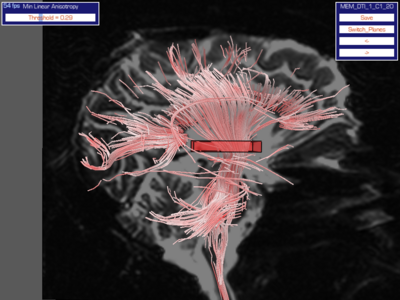
[[Image:Corticospinal Tract ROI 2.png|thumb|400px|center|A single-box ROI that captures the CST as well as several extra streamtubes. This selection must be refined to reject non-CST tubes. See the gallery of images below.]]
# Turn on the b=0 image in mid-sagittal view.
# Turn on the b=0 image in mid-sagittal view.
# In one hemisphere, place a rectangular box that is longer in the L-R and A-P directions than the S-I direction (flat, like a pizza box, aligned in the transverse plane).
# In one hemisphere, place a rectangular box that is longer in the L-R and A-P directions than the S-I direction (flat, like a pizza box, aligned in the transverse plane).
Line 7:
Line 9:
#* below the body of the fornix.
#* below the body of the fornix.
#;The thalamus may be obscured by CSF in a truly mid-sagittal b=0 slice. The box should be placed about in the middle of the S-I extent of the thalamus.
#;The thalamus may be obscured by CSF in a truly mid-sagittal b=0 slice. The box should be placed about in the middle of the S-I extent of the thalamus.
<gallery>
Image:Corticospinal Tract ROI 1.png|Initial placement of the box.
Image:Corticospinal Tract ROI 2.png|Box with streamtubes passing through it.
Image:Corticospinal Tract ROI 3.png|Refined selection with smaller boxes to add or remove streamtubes.
</gallery>
[[Category:Diffusion MRI]]
[[Category:Diffusion MRI]]
Latest revision as of 02:10, 6 March 2009
Here are some loose guidelines for placing the selection boxes to capture the corticospinal tract (CST).
A single-box ROI that captures the CST as well as several extra streamtubes. This selection must be refined to reject non-CST tubes. See the gallery of images below.
Turn on the b=0 image in mid-sagittal view.
In one hemisphere, place a rectangular box that is longer in the L-R and A-P directions than the S-I direction (flat, like a pizza box, aligned in the transverse plane).
Position the box medially in the fiber bundle so that it captures the full extent of the CST as it courses past the thalamus. The box should be situated approximately as follows:
above the mamillary bodies and superior colliculi at the level of the thalamus, but
below the body of the fornix.
The thalamus may be obscured by CSF in a truly mid-sagittal b=0 slice. The box should be placed about in the middle of the S-I extent of the thalamus.
Initial placement of the box.
Box with streamtubes passing through it.
Refined selection with smaller boxes to add or remove streamtubes.

 Initial placement of the box.
 Box with streamtubes passing through it.
 Refined selection with smaller boxes to add or remove streamtubes.
